
In this article, the causes and treatment methods for eyelid entropion of dogs
The owners often find that the dog's lower eyelids are somewhat transverse, but sometimes it is normal. Why does the dog's eyes turn outward? What are its symptoms? How to treat it?
1. Epilosus
Epilosus refers to the partial or all of the eyelid margins turning outward, the eyelid conjunctiva reveals and the formation of rabbit eyes. The lower eyelids are more common, but the upper eyelids are also scarred.
A Saint Bernard dog (detailed introduction) has a rhombus eye, which is manifested as severe lower eyelid envelopment, while the inner corner of the lower eyelid is inverted. The inner and outer corners of the upper eyelid are inverted, secondary corneal stromal ulcers, whole corneal edema, and accompanied by marginal angiogenesis and chronic keratitis.
2, Cause of the disease
1.) Developmental valgus is related to congenital inheritance, and is commonly found in St. Bernard, Bloody, Great Dane, Newfoundland and Bullmastiff. These dogs are often loose with lower eyelids, which may be caused by obvious broad eyelids and lack of lateral contractile muscles, and are also standard characteristics of this breed of dogs.
2.) Acquired eclipse caused by trauma or chronic inflammation, scar formation, fatigue, and sagging of the eyelid fissures and lesion nerve damage in elderly dogs can all cause this disease.
3. Symptoms
The lower eyelid is entropic, the eyelid conjunctiva and bulbous conjunctiva are exposed, red or dark red. Due to the long-term exposure of the conjunctiva, it causes conjunctiva inflammation and tears, and the hair in the eyes is wet.
4. Treatment
For cases where lower eyelid entropion is not severe, dogs with chronic tear overflow do not need surgery. Antibiotics and corticosteroid eye drops can be used to spot the eyes (dexamethasone, etc.) to reduce local irritation and prevent infection. If the secretions are abnormally increased and chronic conjunctivitis and blepharospasm occurs, surgical correction is suitable. There are many ways to correct this disease, but generally use warton-Jones lesion formation surgery, which is V-Y technology. A V-shaped skin incision with deep large subcutaneous tissue is made at 2-3mm below the lid edge of the eccentric lower eyelid, and the V-shaped base should be wider than the eccentric part of the lid edge. Then the subcutaneous tissue is separated upward from the tip of the V-shaped incision and gradually free the triangular flap. Then perform appropriate seismic separation under the skin of the wound edges on both sides, and do nodules suture from the V-shaped tip upwards. Move the flap upwards while suturing until the eclipse of the lower eyelid eyelid eyelid is restored to its original state and corrected. Finally, the nodules suture the remaining skin incision, which is to change the original V-line incision into a Y-shaped shape. The operation is often done with No. 4 or No. 7 silk threads, keep the needle distance of 2 mm, and remove the sutures 10-14 days after the operation.
5. Postoperative care
After the operation, antibiotic eye drops or eye ointment should be used, 3-4 times a day for 5-7 days. Eliminate the symptoms of conjunctivitis or keratitis secondary to eyelid eclipse; pay attention to surgical injuries caused by scratching or friction of animals.
Recommend News
-
Xiaomengmao was full of grievance when she was given the first time she was vacc
-
What should the kitten feed him when he gets angry? What is the correct feeding
-
How to train your cat to become a social expert? Cultivate the social behavior o
-
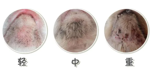
The cat s chin is black, what should I do if the cat s chin is black?